
Classification of Human Brain: Structure, Stages, and Biology
Have you ever wondered how the brain controls your body every day? Understanding the classification of human brain anatomy makes this complex organ easier to study. Millions of neurons work together to control memory, movement, emotions, thinking, and vital body functions.
Neuroscientists classify the brain using structural, functional, and developmental models. These systems help researchers understand how different regions work together. They also support the diagnosis and treatment of neurological disorders. For a reliable introduction to brain structure and function, explore the National Institute of Neurological Disorders and Stroke (NINDS) Brain Basics.
The brain can be studied in several ways. We can examine its major anatomical divisions, follow its development from early embryonic stages, or study the functions of individual regions. We can also place humans within the broader biological classification system. For students, educational psychology theories can also provide useful context for understanding how learning and cognitive processes relate to education.
This guide explains the major approaches to brain classification. It also covers the seven stages of brain development, the three major brain divisions, seven important brain parts, and the biological classification of humans. This makes the topic useful for students, exam preparation, and anyone interested in basic neuroscience.
What is classification in the brain?
Classification in the brain means organizing neural structures according to their location, development, structure, and function. Instead of treating the brain as one uniform organ, scientists divide it into related regions. Each region performs specific tasks while working with other areas.
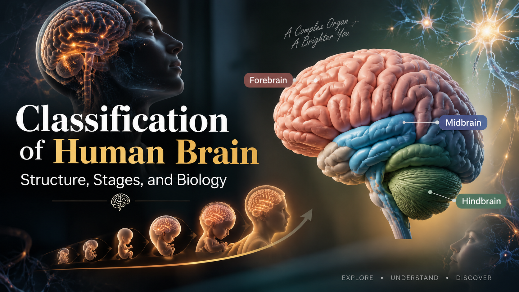
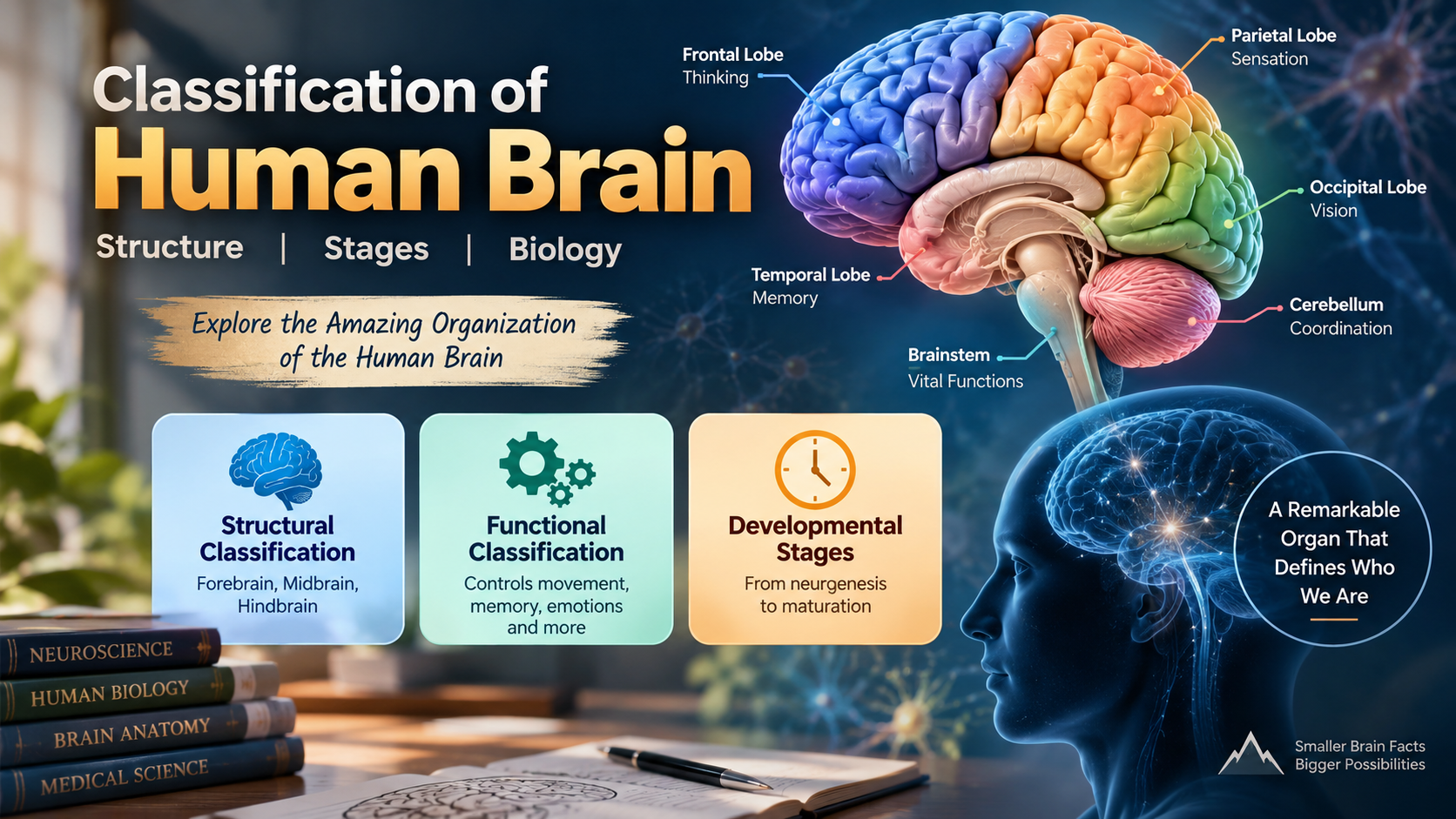
The main structural classification of human brain anatomy commonly includes the forebrain, midbrain, and hindbrain. These divisions help students understand the broad organization of the central nervous system.
Scientists can also classify brain regions by their functions. Some areas process sensory information, while others control movement, emotions, memory, or higher cognitive activities. Developmental classification examines how these structures form during embryonic growth.
These classification systems are useful in medicine and research. Doctors use brain anatomy to interpret imaging results and understand injuries. Researchers also use these divisions to study neurological conditions and changes in brain function.
Anatomical Versus Functional Categorization
Anatomical classification organizes brain structures according to their physical location, shape, and relationships with nearby regions. Important anatomical divisions include the cerebral hemispheres, lobes, brainstem, cerebellum, and other internal structures.
Functional classification focuses on what each region does. For example, the occipital lobe is strongly associated with visual processing, while the frontal lobe supports planning, decision-making, and voluntary movement.
These approaches are not separate in practice. Structure and function are closely connected. A particular brain region has physical features that support its specialized activities.
Understanding both systems also helps explain the effects of brain damage. Injury to one area may affect movement, speech, memory, vision, or other abilities. However, brain functions often involve networks rather than a single isolated region. Therefore, modern neuroscience combines anatomical and functional information when studying brain organization.
What are the 7 stages of the brain?
The seven stages of brain development describe major biological processes involved in building and refining the nervous system. These stages include neurogenesis, cell migration, differentiation, synaptogenesis, synaptic pruning, myelination, and structural maturation.
Neurogenesis produces new neurons from developing neural cells. During cell migration, immature neurons move toward their appropriate locations. Differentiation then allows these cells to develop specialized characteristics and functions.
Synaptogenesis creates connections between neurons. The developing brain initially forms many connections, creating flexible neural networks. Synaptic pruning later removes weaker or less-used connections. This process helps make neural communication more efficient.
Myelination adds protective myelin around many nerve fibers. This insulation improves the speed and efficiency of electrical signaling. Finally, structural maturation continues as brain networks become more organized and specialized.
These processes do not occur as perfectly separate steps. They overlap and continue at different rates across brain regions. Brain development also continues beyond childhood, particularly in areas involved in planning, decision-making, and self-control. These differences also help explain why personalized learning in education can account for variations in how individuals learn and develop.
Synaptic Pruning and Efficient Cognition
During childhood and adolescence, the developing brain creates many more synaptic connections than it eventually needs. Synapses are communication points where neurons exchange signals. This early abundance gives the brain flexibility while neural circuits develop.
Synaptic pruning removes connections that are weak, inefficient, or used less frequently. At the same time, frequently used pathways can become stronger and more efficient. This process helps refine neural networks as a person gains experience.
Pruning does not simply mean that the brain loses connections. Instead, it helps organize existing networks and improve communication between neurons. This refinement supports increasingly complex cognitive abilities.
The process continues throughout development, although its timing differs across brain regions. Areas involved in higher-level functions can mature later than some sensory and motor regions.
For students, the key idea is simple: synaptic pruning helps the developing brain become more efficient. Experience, learning, and repeated activity can influence which neural pathways become stronger and which connections are reduced. Applying this understanding to best study techniques backed by science can help students connect repeated learning activities with effective study habits.
What are the 3 major parts of the brain and their functions class 10?
For Class 10 biology, the brain is commonly divided into three major regions: the forebrain, midbrain, and hindbrain. This simple classification helps students understand the basic organization and functions of the human nervous system.
The forebrain is involved in conscious activities, sensory interpretation, memory, thinking, and voluntary actions. Its largest structure is the cerebrum, which supports many higher mental functions.
The midbrain connects parts of the brain and participates in important reflex responses. It helps coordinate certain visual and auditory reflexes.
The hindbrain includes the cerebellum, pons, and medulla oblongata. The cerebellum helps coordinate movement, posture, and balance. The pons participates in communication between brain regions and contributes to breathing regulation. The medulla controls several involuntary functions, including breathing, heart activity, and blood pressure.
For Class 10 exams, remember the basic division and the major functions of each region. This provides a clear foundation for understanding the classification of human brain. Students can also strengthen their preparation by using study tips for exam preparation alongside this foundational biology knowledge.
Key Examination Takeaways for Forebrain and Hindbrain
For examinations, focus on the major differences between the forebrain and hindbrain. The forebrain is strongly associated with conscious activities, sensory interpretation, thinking, memory, and voluntary actions.
The cerebrum forms the largest part of the forebrain. It receives and interprets sensory information and supports complex activities such as reasoning, learning, and decision-making.
The hindbrain performs several essential coordination and automatic functions. Its cerebellum helps maintain balance, posture, and coordination during movement. The medulla oblongata controls important involuntary activities, including breathing, heart rate, and blood pressure.
A simple way to remember the difference is to connect the forebrain with higher mental and voluntary functions. Associate the hindbrain with coordination and vital automatic functions.
Students should also avoid treating every brain function as belonging to only one structure. Many activities involve several connected regions. For school-level questions, however, learning the primary functions of each division provides a useful and accurate framework.
What are the 7 parts of the brain and their functions?
A useful overview of brain anatomy identifies seven major structures: the cerebrum, cerebellum, diencephalon, midbrain, pons, medulla oblongata, and limbic system. Each contributes to different aspects of nervous system activity.
The cerebrum supports conscious thought, sensory processing, memory, language, and voluntary movement. The cerebellum helps coordinate movement, balance, posture, and motor learning.
The diencephalon contains structures such as the thalamus and hypothalamus. The thalamus relays much sensory information, while the hypothalamus helps regulate temperature, hunger, thirst, and endocrine activity.
The midbrain participates in visual and auditory reflexes. The pons helps connect different brain regions and contributes to breathing and sleep-related functions. The medulla oblongata controls several essential automatic processes, including breathing and cardiovascular regulation.
The limbic system includes interconnected structures involved in emotion, motivation, learning, and memory. These seven categories provide a practical overview, although modern neuroscience recognizes many additional structures and functional networks.
The Diencephalon’s Critical Role as Relay Station
The diencephalon is an important region located deep within the brain. It includes the thalamus, hypothalamus, and several other structures. Two of its best-known components have especially important roles in sensory processing and internal regulation.
The thalamus acts as a major relay center for sensory information. It receives signals from many parts of the nervous system and sends much of that information toward appropriate areas of the cerebral cortex. This helps the brain organize and interpret incoming sensory information.
The hypothalamus has a different but equally important role. It helps maintain homeostasis, which means keeping internal conditions within suitable ranges. It contributes to temperature regulation, hunger, thirst, sleep, and hormonal control.
The hypothalamus also communicates closely with the endocrine system through its relationship with the pituitary gland. This allows the nervous and hormonal systems to coordinate many body processes.
Therefore, the diencephalon is more than a simple relay station. It connects sensory processing with essential systems that maintain the body’s internal balance.
What are the 7 biological classifications?
The seven traditional biological classifications are Kingdom, Phylum, Class, Order, Family, Genus, and Species. These taxonomic ranks organize living organisms from broad groups to increasingly specific categories.
Kingdom represents a broad biological grouping. Phylum divides organisms according to major structural and evolutionary characteristics. Class provides a more specific grouping, followed by Order and Family. Genus groups closely related organisms, while Species represents the most specific standard rank in this seven-level system.
Humans can be classified as follows:
- Kingdom: Animalia
- Phylum: Chordata
- Class: Mammalia
- Order: Primates
- Family: Hominidae
- Genus: Homo
- Species: Homo sapiens
This classification places humans within the animal kingdom and shows their evolutionary relationships with other organisms. It is important to distinguish this biological taxonomy from the anatomical classification of human brain. One classifies organisms, while the other organizes brain structures.
Modern biology may also use additional ranks and molecular evidence. Therefore, the seven-rank system is best understood as a traditional framework for organizing biological diversity.
Evolutionary Context of Human Taxonomy
Human taxonomy places our species within Kingdom Animalia, Phylum Chordata, Class Mammalia, and Order Primates. This classification reflects shared evolutionary characteristics with other animals and, more specifically, with other primates.
Humans belong to the family Hominidae and the genus Homo. Our species is Homo sapiens. These categories help scientists describe evolutionary relationships using a standardized biological system.
Humans share many characteristics with mammals, including specialized nervous systems, internal temperature regulation, and particular reproductive features. Our placement within Primates reflects shared traits with other primates, including grasping hands and complex social behavior.
Over evolutionary time, the human nervous system developed increasingly complex structures and networks. The cerebral cortex became highly developed, supporting advanced communication, planning, learning, and flexible problem-solving.
However, human evolution should not be described as a simple progression from a primitive brain to a perfect one. Evolution produces adaptations in response to changing environments and pressures. Understanding human taxonomy therefore provides context for studying how our distinctive brain features developed within the broader history of life.
Frequently Asked Questions
What is the difference between grey matter and white matter?
Grey matter contains neuronal cell bodies, dendrites, and synapses that process information. White matter contains myelinated axons that transmit signals between brain regions. Both are essential to the classification of human brain structures.
Which part of the brain controls voluntary movement?
The primary motor cortex in the frontal lobe controls voluntary movement. It sends signals through spinal pathways to skeletal muscles. The cerebellum and basal ganglia help refine and coordinate these movements.
How does the nervous system protect the brain from injury?
The brain is protected by the skull, three layers of meninges, and cerebrospinal fluid. The blood-brain barrier also limits harmful substances and pathogens from reaching sensitive neural tissue.
Why is the human cerebral cortex so heavily folded?
The cerebral cortex contains folds called sulci and gyri that increase its surface area within the limited skull space. This arrangement supports more neurons and complex functions such as reasoning, language, and memory.
Conclusion
Mastering the classification of human brain structures clarifies how biological form directly supports neurological function. By studying the brain through anatomical divisions, embryonic stages, and vital regional responsibilities, we transform an intimidating organ into an accessible biological network. From the reflex coordination managed by the hindbrain to the abstract deliberations housed in the cerebral cortex, every neural division performs a specialized job that sustains our daily lives. Furthermore, framing this neurological marvel within the broader tree of biological taxonomy reminds us of our evolutionary roots.
As modern neuroimaging advances, these anatomical boundaries become even more valuable for diagnosing cognitive illnesses and developing targeted brain therapies. Whether you are analyzing these concepts for an upcoming academic exam or enriching your personal understanding of human physiology, this foundational knowledge highlights how dynamic our central control center truly is. Continue exploring neuroscience literature, test your recall on these major structural divisions, and apply these valuable insights to your ongoing scientific studies.